7 september 2014: Bron: Ann Oncol. 2014;25(8):1536-1543.
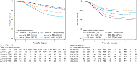
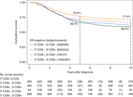
Aanwezigheid van CD8 positieve expressie plus T-cellen in de tumor en beenmerg wijzen op een significant verlaagd risico op overlijden aan borstkanker. Percentages van 21% tot soms 57% minder risico op overlijden zijn gevonden, afhankelijk van de plaats van de tumoren. Dit gaat op voor zowel oestrogeen positieve vormen van borstkanker met HER2-positieve receptor (ER-positief) als voor borstkanker met ER-negatieve vormen van borstkanker.
Dit blijkt uit een grote meta analyse van 4 gerandomiseerde studies bij totaal 12.439 patiënten met borstkanker. CD8 expressie en T-cellen zijn vantevoren te meten. Bv. via een biomoleculair profile onderzoek.
Het mooie van deze studie is dat CD8 positieve expressie als T-cel infiltratie blijkbaar geen onderscheid maakt tussen welke vorm van borstkanker wat betreft hormoonstatus (ER-pos of ER-neg) enz.. De onderzoekers stellen zelfs dat borstkanker blijkbaar een meervoudige moleculair gerelateerde ziekte is en geen enkele ziekte op zichzelf.

Deze studie van 12 439 vrouwen met borstkanker is de grootste evaluatie van T-cellen als een tumor marker in data van patiënten met borstkanker.
Hieruit blijkt dat de aanwezigheid van CD8 + T-cellen in ER-negatieve borsttumoren is geassocieerd met een vermindering van het relatieve risico op sterven aan borstkanker tussen de 57% en 21% afhankelijk van hun locatie (iT, Stroma of beide) en voor iT-CD8 + T-cellen vonden we een vermindering van 27% in het risico op overlijden aan borstkanker met ER-positieve HER2-positieve tumoren.
We namen een groot aantal goed gekarakteriseerde patiënten in deze studie en daarmee zijn onze conclusies ook statistisch onderbouwd. Daarnaast konden wij borstkanker evalueren als zijnde een groep van ziekten met moleculaire subtypes in plaats van een enkele entiteit. Aldus de onderzoekers in hun studieverslag
(This study of 12 439 women with breast cancer is the largest evaluation of T cells as a tumour marker in breast cancer to date. It shows that the presence of CD8+ T cells in ER-negative breast tumours is associated with a reduction in the relative hazard of dying from breast cancer of between 57% and 21% depending on their location (iT, S or both) and, for iT-CD8+ T cells, with a 27% reduction in the hazard of dying from breast cancer in ER-positive HER2-positive tumours.)
We included a large number of well-characterised patients in this study and, therefore, our conclusions are statistically robust. In addition, we have been able to evaluate breast cancer as a group of related diseases (molecular subtypes) rather than a single entity.

Zie hier het volledige studierapport: Association Between CD8+ T-cell Infiltration and Breast Cancer Survival in 12 439 Patients dat gratis is in te zien.
Hier het abstract van de studie.
The presence of CD8+ T cells in breast cancer is associated with a significant reduction in the relative risk of death from disease in both the ER-negative and the ER-positive HER2-positive subtypes. Tumour lymphocytic infiltration may improve risk stratification in breast cancer patients classified into these subtypes.
Source:
-
Ann Oncol (2014) 25 (8): 1536-1543. doi: 10.1093/annonc/mdu191
Association between CD8+ T-cell infiltration and breast cancer survival in 12 439 patients
- H. R. Ali1,2,3,
- E. Provenzano1,3,
- S.-J. Dawson1,3,4,
- F. M. Blows4,
- B. Liu1,3,4,
- M. Shah3,4,5,
- H. M. Earl3,4,
- C. J. Poole6,
- L. Hiller6,
- J. A. Dunn6,
- S. J. Bowden7,
- C. Twelves8,
- J. M. S. Bartlett9,10,
- S. M. A. Mahmoud11,†,
- E. Rakha11,
- I. O. Ellis11,
- S. Liu12,
- D. Gao12,
- T. O. Nielsen12,
- P. D. P. Pharoah3,4,5 and
- C. Caldas1,3,4,*
+ Author Affiliations
- ↵*Correspondence to: Dr Carlos Caldas, Cancer Research UK Cambridge Institute, University of Cambridge, Li Ka Shing Centre, Robinson Way, Cambridge CB2 0RE, UK. Tel: +44-1223-769650; E-mail: carlos.caldas@cruk.cam.ac.uk
- Received May 5, 2014.
- Revision received May 8, 2014.
- Accepted May 8, 2014.
Abstract
Background T-cell infiltration in estrogen receptor (ER)-negative breast tumours has been associated with longer survival. To investigate this association and the potential of tumour T-cell infiltration as a prognostic and predictive marker, we have conducted the largest study of T cells in breast cancer to date.
Patients and methods Four studies totalling 12 439 patients were used for this work. Cytotoxic (CD8+) and regulatory (forkhead box protein 3, FOXP3+) T cells were quantified using immunohistochemistry (IHC). IHC for CD8 was conducted using available material from all four studies (8978 samples) and for FOXP3 from three studies (5239 samples)-multiple imputation was used to resolve missing data from the remaining patients. Cox regression was used to test for associations with breast cancer-specific survival.
Results In ER-negative tumours [triple-negative breast cancer and human epidermal growth factor receptor 2 (human epidermal growth factor receptor 2 (HER2) positive)], presence of CD8+ T cells within the tumour was associated with a 28% [95% confidence interval (CI) 16% to 38%] reduction in the hazard of breast cancer-specific mortality, and CD8+ T cells within the stroma with a 21% (95% CI 7% to 33%) reduction in hazard. In ER-positive HER2-positive tumours, CD8+ T cells within the tumour were associated with a 27% (95% CI 4% to 44%) reduction in hazard. In ER-negative disease, there was evidence for greater benefit from anthracyclines in the National Epirubicin Adjuvant Trial in patients with CD8+ tumours [hazard ratio (HR) = 0.54; 95% CI 0.37−0.79] versus CD8−negative tumours (HR = 0.87; 95% CI 0.55–1.38). The difference in effect between these subgroups was significant when limited to cases with complete data (Pheterogeneity = 0.04) and approached significance in imputed data (Pheterogeneity = 0.1).
Conclusions The presence of CD8+ T cells in breast cancer is associated with a significant reduction in the relative risk of death from disease in both the ER-negative [supplementary Figure S1, available at Annals of Oncology online] and the ER-positive HER2-positive subtypes. Tumour lymphocytic infiltration may improve risk stratification in breast cancer patients classified into these subtypes.
NEAT ClinicalTrials.gov NCT00003577.
Gerelateerde artikelen
- Vloeibare biopsie (bloedtest) op zoek naar circulerend tumor-DNA (ctDNA) is een steeds vaker gebruikte methode bij de preventie, diagnose en behandelingen van borstkanker. Hier een uitleg aan de hand van de literatuur.
- Clairity, een met AI Kunstmatige Intelligentie ontwikkeld hulpmiddel, ontdekt veel eerder ontstaan van borstkanker in mammografiebeelden en is nu door de FDA officieel erkent als hulpmiddel bij een mammografie
- bloedtest na behandeling van borstkanker waarbij resistentie optreedt traceert HER2 mutaties die soms ontstaan tijdens de behandeling met Kinaseremmers en hormoontherapie copy 1
- BRIP1 mutatie geeft uitstekende respons op olaparib bij uitgezaaide HR-positieve, HER2-negatieve borstkanker blijkt uit casestudie
- Tumorcellen in bloed bij patienten met operabele beginnende borstkanker voorspellen door betere resultaten noodzaak van radiotherapie - bestraling in vergelijking met geen tumorcellen in bloed.
- Tumorcellen in bloed bij patienten met operabele beginnende borstkanker voorspellen door betere resultaten noodzaak van radiotherapie - bestraling in vergelijking met geen tumorcellen in bloed. copy 1
- DNA onderzoek van botweefsel (bone-in-culture array - BICA) ontdekt en adviseert effectieve behandelingen voor botmetastases van borstkanker
- Amastest lijkt goede en betrouwbaar alternatieve diagnose techniek, maar studies tonen grote verschillen
- Borstkanker: Voorspellende biomoleculaire markers en DNA mutaties bij borstkanker en uitgezaaide borstkanker: een overzichtstudie vanuit het Preagnant netwerk copy 1
- Borstkanker: CD8 positieve expressie plus T-cel infiltratie in tumor en beenmerg voorspellen als markers een significant verlaagd risico - 21 tot 57 procent - op overlijden aan borstkanker, zowel bij ER-HER2 pos als ER neg. vormen van borstkanker copy 2
- Borstkanker: Enzalutamide - Xtandi geeft spectaculaire resultaten bij gevorderde triple negatieve borstkanker (stadium 4) met ook een positieve expressie van de hormoonreceptor. copy 2
- Borstkanker: PKI3CA enzym expressie bepalend voor verwacht resultaat van chemo behandeling bij borstkanker en is gerelateerd aan HER2 status en hormoonstatus copy 1 copy 1
- Beenmergpunctie: Losse kankercellen in beenmerg voorspellen de recidiefkansen voor vrouwen met borstkanker met aangetaste lymfklieren ongeacht welke behandeling dan ook.
- Biopt uit schildwachtklier bij borstkanker veroorzaakt niet significant uitzaaiingen in lymfklieren of elders in lichaam dan zonder biopt, aldus gerandomiseerde studie bij 2502 vrouwen.
- Bloedtest voorspelt kans en duur op overleving van patiënten met uitgezaaide borstkanker.
- CD8 positieve expressie plus T-cel infiltratie in tumor en beenmerg voorspellen als markers een significant verlaagd risico - 21 tot 57 procent - op overlijden aan borstkanker, zowel bij ER-HER2 pos als ER neg. vormen van borstkanker copy 1
- Diagnose: Wanneer kanker in tweede borst vroeg wordt ontdekt en tijdig wordt behandeld is de kans op overlijden 27% tot 47% kleiner t.o.v. latere ontdekking van een recidief.
- Genentest Oncotype DX bij patienten met hormoongevoelige borstkanker (ER pos. Her2 neg.) heeft een directe relatie met wel of geen chemo en risico op overlijden en zou standaard ingevoerd moeten worden
- HER-2-neu status verandert bij ca. 26% van de vrouwen met borstkanker door gebruik van Tamoxifen en Letrazole - Femara en verandert Her2-Neu expressie van negatief naar positief.
- Hoe langer het duurt voordat borstdichtheid afneemt bij ouder wordende vrouwen hoe groter het risico op ontstaan van borstkanker.
- MRI-scan naast het standaard bevolkingsonderzoek voor borstkanker is gevoeliger en kan vrouwen met dicht borstweefsel helpen tumoren te ontdekken maar geeft vaker vals alarm copy 1
- MRI scan, Ultrasound, Petscan en scintimammography zijn onvoldoende accuraat in vaststellen van borstkanker in vergelijking met een biopt
- Nieuwe diagnosetechniek met oplichtende contrastvloeistof superieur aan standaard mammografie
- Mammaprint - genentest bewijst waarde voor borstkanker en kan ook uitgevoerd worden op bewaard tumorweefsel.
- Oestrogeen - ER, progesteron- PR en HER2 receptor expressie laat groot verschil (42 procent) zien tussen primaire tumor en uitzaaiingen in de hersenen bij borstkanker en heeft groot effect op behandeling en overleving
- Opsporing borstkanker via PET scan geeft ca. 90% zekerheid bij diagnose van eventueel recidief van borstkanker.
- Perifere circulerende tumorlymfocyten (pCTL) in bloed blijken alleen voorspellend te zijn voor HER2-positieve ziekte bij borstkankerpatiënten die werden behandeld met anti-HER2 medicijnen.
- PIK3CA mutatie geeft minder therapeutisch effect van anti-HER2 medicijnen zoals lapatinib, trastuzumab - herceptin en de combinatiebehandeling van lapatinib en trastuzumab - herceptin samen
- Receptorstatus van primaire tumor bij vrouwen met uitgezaaide borstkanker verschilt gemiddeld 31 procent (range 20 tot 65 procent bij HER2+ subtype) met die van de receptorstatus van de uitzaaiingstumor copy 1
- Schildwachtklier methode bij borstkanker, hoe werkt dat?
- Spontane genezingen: 22 procent van beginnende borstkanker zou spontaan genezen suggereert een groot langjarige bevolkingsonderzoek onder 100.000 Noorse vrouwen naar effecten en noodzakelijkheid van mammografie.
- Ultra sound diagnostiek aanvullend op mammografie diagnosteert nog eens 15 procent extra van verdacht weefsel. Blijkt uit langjarige gerandomiseerde studie.
- Diagnose en oorzaken van borstkanker: een overzicht van artikelen en recente ontwikkelingen.


Plaats een reactie ...
Reageer op "Borstkanker: CD8 positieve expressie plus T-cel infiltratie in tumor en beenmerg voorspellen als markers een significant verlaagd risico - 21 tot 57 procent - op overlijden aan borstkanker, zowel bij ER-HER2 pos als ER neg. vormen van borstkanker copy 2"