Mocht u kanker-actueel de moeite waard vinden en ons willen ondersteunen om kanker-actueel online te houden dan kunt u ons machtigen voor een periodieke donatie via donaties: https://kanker-actueel.nl/NL/donaties.html of doneer al of niet anoniem op - rekeningnummer NL79 RABO 0372931138 t.n.v. Stichting Gezondheid Actueel in Amersfoort. Onze IBANcode is NL79 RABO 0372 9311 38
Elk bedrag is welkom. En we zijn een ANBI instelling dus uw donatie of gift is in principe aftrekbaar voor de belasting.
30 april 2017: Bron: Nat Commun. 2017 Apr 21;8:15045. doi: 10.1038/ncomms15045
Danusertib zou het beste medicijn zijn om uitzaaiingen in de botten o.a. vanuit borstkanker te behandelen. Dat stellen Amerikaanse onderzoekers van het Baylor College of Medicine en andere instituten in een publicatie van de resultaten uit een dierstudie in Nature Communications.
Zij hebben een nieuwe laboratoriumtechniek ontwikkeld die de effectiviteit van bepaalde medicijnen voor uitzaaiingen in de botten, zoals in deze studie vanuit borstkanker snel kunnen testen buiten de patiënt om. Zij noemen deze nieuwe techniek "BICA - bone-in-culture array". Daarmee kunnen ze snel en zonder de patiënt extra te belasten de beste opties voor een behandeling ontdekken. In deze dierstudie met muizen met borstkankeruitzaaiingen ontdekten zij dat met name danusertib, een zogeheten Aurora kinase remmer, het beste medicijn bleek om botuitzaaiingen vanuit borstkanker te behandelen. Maar ook bij andere botmetastasen bleek danusertib effectief.
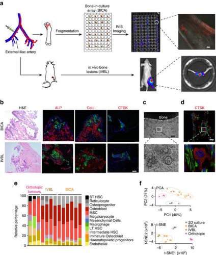
Hier een schema hoe de BICA werkt: (tekst gaat onder schema verder):
(a) Schematic of IIA injection-based establishment of BICA and IVBL. Luciferase/fluorescence protein-tagged cancer cells are inoculated into the external iliac artery of mice. The injected animals will either be killed for BICA or left alive to give rise to IVBL. For BICA set-up, femur and tibia bones are extracted. Epiphysis and metaphysis are segmented into pieces (1–2 mm in diameter), which will then be arranged into low-attachment 96-well plates with cell culture media. Cancer cells can be traced using confocal microscopy and bioluminescence imaging. Scale bar in upper right panel, 100 μm. (b) Haematoxylin and eosin, and fluorescence staining of osteoblast (ALP and Col-I) and osteoclast (CTSK) markers to characterize the microenvironment niche of MCF-7 cells (green) in BICA and IVBL. The quantification is shown in Supplementary Fig. 1d. Scale bar, 25 μm. (c) Inverted microscope images of a bone fragment in BICA and haematopoietic cells staying around it. The white dotted line indicates the border of bone fragment. Scale bar, 100 μm. (d) Fluorescence staining in BICA indicated CTSK+ (red) osteoclasts and human-specific Vimentin+ (green) SCP28 cells on a BICA fragment of 5 weeks culture without treatment of M-CSF and RANKL. Scale bar, 25 μm. (e) Stromal components of MCF-7 orthotopic tumours, IVBL and BICA predicted by CYBERSORT algorithm to mouse sequences in RNA-seq results. Each column represents one tumour (orthotopic), one bone (IVBL) or pooled bone segments from one animal (BICA). ST HSC, short-term haematopoietic stem cells; LT HSC, long-term haematopoietic stem cells. (f) Principle component analysis (PCA) of human sequences in the RNA-seq results of the indicated MCF-7 specimens. Upper panel: PCA based on the top 100 genes with highest average expression across all samples. PC1/2, principle components 1/2. Lower panel: t-Distributed Stochastic Neighbor Embedding (t-SNE) analysis17.
"Voor een aantal patiënten met borstkanker zijn uitzaaiingen het probleem nadat de primaire tumor is geëlimineerd," aldus een van de auteurs Xiang Zhang, PhD, Associate Professor of Molecular and Cellular Biology at the Lester and Sue Smith Breast Center at Baylor.
"Metastasen - uitzaaïngen hebben echter de neiging om anders te reageren dan de primaire tumor op een behandeling, onder meer doordat deze vaak voorkomen in een ander orgaan met een andere micro-omgeving." "Tot nu toe is er geen effectief experimenteel platform gevonden om uitgezaaide tumoren te bestuderen in hun nieuwe micro-omgeving."
"We hebben een experimenteel systeem gecreëerd waarin we de interacties tussen kankercellen en botcellen kunnen nabootsen, omdat bot de plek is waar borstkanker en veel andere kankers zich meestal naar verspreiden," zegt Dr. Zhang. "We hebben een systeem ontwikkeld waarmee we veel verschillende medicijneracties tegelijk kunnen testen om de juiste behandeling te ontdekken die selectief effectief is voor uitzaaiende kankercellen en het bij effect op het bot zelf te minimaliseren."
Om de interacties tussen metastatische borstkankercellen en botcellen in een levend systeem in het laboratorium na te bootsen, ontwikkelden Dr. Zhang en zijn collega's een botmetastasemodel, dat zij een "bone-in culture array" noemen. De wetenschappers hebben vastgesteld dat de bone-in-culture array de micro-milieu karakteristieken van botmetastases behoudt in levende dierenmodellen. Ook zagen zij dat de kankercellen het DNA- en receptorenexpressie profiel behielden evenals het groeipatroon en de respons op medicijnen.
Met behulp van de bone-in-culture array, ontdekten de onderzoekers dat het medicijn danusertib, een zogeheten pan-aurora kinaseremmer, het beste werkte bij botmetastases. Zij vonden ook dat andere medicijnen de groei van langzame groeiende kankercellen in het bot juist stimuleerden.
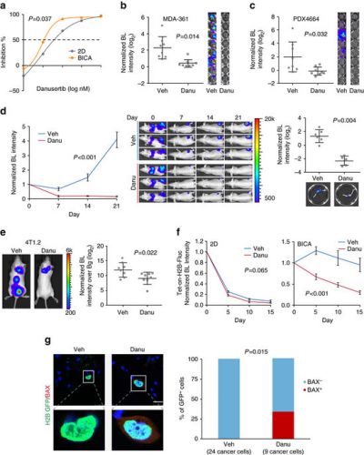
Hier een beeld van hoe danusertib effectiviteit laat zien in hun studie: (tekst gaat onder grafiek verder)
(a) Dose responsive curves of MCF-7 cells to danusertib in 2D culture or BICA. For 2D cultures, six technical replicates were included for each drug concentration. For BICA, N=6 bone fragments for each drug concentration. Samples are under corresponding treatment for 3 weeks and measured by bioluminescence (BL). P values are determined by repeated measures analysis of variance (ANOVA) tests. (b,c) The effects of danusertib (100 nM) on MDA-MB-361 cells (b) and PDX4664 cells (c) in BICA. N=8 bone fragments in each group. Samples are under corresponding treatment for 3 weeks. Veh, vehicle; Danu, danusertib. P values are determined by Student’s t-test (two-tailed) with Welch correction. Error bars: s.d. (d) The effects of danusertib on IIA-injected MCF-7 bone lesions. Left: growth curves as measured by in vivo BL imaging. N=6 and 5 athymic nude mice for the two groups, respectively. P values are determined by repeated measures ANOVA tests on the growth curves. Error bars: s.e.m. Middle: representative BL pictures show bone lesion arising with or without danusertib treatment. Right: quantification of BL intensity after hindlimbs are extracted. P values were determined by two-tailed unpaired Mann–Whitney U-test. Error bars: s.d. (e) The effects of danusertib on spontaneous bone metastasis of 4T1.2 model. The treatment of danusertib started after orthotopic tumour resection. Metastasis was quantified 12 days after the surgery, as shown in the dot plot. N=8 Balb/c mice in each experimental group. Bg, background. The P value was determined by t-test (two-tailed). Error bars: s.d. (f) The effects of danusertib on dormant MCF-7 cells in BICA as measured by Tet-on inducible H2B-Fluc/GFP system. N=6 and 8 bone fragments in each group, respectively. P values are determined by repeated measures ANOVA tests. Error bars: s.e.m. (g) Immunofluorescence staining and quantification of GFP (green) in conjunction with BAX (red) on MCF-7 cells in BICA with 1 week of treatment after IIA injection. N=8 bone fragments in each group. Numbers in parentheses indicate the quantity of cancer cells examined. Scale bar, 25 μm. The P value was determined by Fisher’s exact test.
Volgens de onderzoekers kan hun BICA - bone-in-culture array ook het effect bepalen bij andere botuitzaaiingen vanuit andere primaire tumoren. Zij probeerden al enkele andere vormen van botuitzaaiingen en ook daarbij bleek danusertib het effectiefste medicijn te zijn.
Het is natuurlijk nog een dierstudie en er zal wel enkele jaren overheen gaan voor dit in de praktijk gebracht zal gaan worden. Aan de andere kant danusertib wordt al veel onderzocht en zijn ook al enkele studieresultaten van bekend. Echter niet alleen voor botmetastases maar bij patienten met vergevorderde kanker zoals o.a. deze studie: Efficacy and safety of biweekly i.v. administrations of the Aurora kinase inhibitor danusertib hydrochloride in independent cohorts of patients with advanced or metastatic breast, ovarian, colorectal, pancreatic, small-cell and non-small-cell lung cancer: a multi-tumour, multi-institutional phase II study.
Of deze studie waarin kinase remmers, waaronder danusertib zijn getest bij botkanker - Multiple myeloma (Kahler): Kinase inhibitors as potential agents in the treatment of multiple myeloma
Dus het medicijn tegen botuitzaaiingen dat uit deze BICA test kwam is al wel beschikbaar. Misschien als u aleen botuitzaaiingen hebt eens bespreken met uw behandelend arts of danusertib beschikbaar is?
Het volledige studierapport: https://www.nature.com/articles/ncomms15045#s1 is gratis in te zien. Hier het abstract van de studie:
The drug danusertib, an investigational pan-aurora kinase inhibitor, preferentially inhibits bone metastasis proves BICA - bone-in-culture array
Bone-in-culture array as a platform to model early-stage bone metastases and discover anti-metastasis therapies
- Nature Communications 8, Article number: 15045 (2017)
- doi:10.1038/ncomms15045
- Download Citation
- Received:
- Accepted:
- Published online:
Abstract
The majority of breast cancer models for drug discovery are based on orthotopic or subcutaneous tumours. Therapeutic responses of metastases, especially microscopic metastases, are likely to differ from these tumours due to distinct cancer-microenvironment crosstalk in distant organs. Here, to recapitulate such differences, we established an ex vivo bone metastasis model, termed bone-in-culture array or BICA, by fragmenting mouse bones preloaded with breast cancer cells via intra-iliac artery injection. Cancer cells in BICA maintain features of in vivo bone micrometastases regarding the microenvironmental niche, gene expression profile, metastatic growth kinetics and therapeutic responses. Through a proof-of-principle drug screening using BICA, we found that danusertib, an inhibitor of the Aurora kinase family, preferentially inhibits bone micrometastases. In contrast, certain histone methyltransferase inhibitors stimulate metastatic outgrowth of indolent cancer cells, specifically in the bone. Thus, BICA can be used to investigate mechanisms involved in bone colonization and to rapidly test drug efficacies on bone micrometastases.
Discussion:
In the current study BICA was primarily used to model pre-osteolytic micrometastases, although we also demonstrated its potential to investigate the onset of osteoclast activation. Perhaps because of limited bone surface area and insufficient monocyte supply, the ability of BICA to model full-fledged vicious cycle is limited. The pre-osteolytic stage corresponds to the phase when adjuvant therapies are applied in the clinic. Although adjuvant therapies are intended to target micrometastases in distant organs, the decision and choice of specific treatments has to be made based on features of primary tumours. In this study we provided three specific examples of how cancer cells in a different context may respond differently to certain drugs. While danusertib exhibited enhanced efficacies on cancer cells interacting with bone, two HMT inhibitors unexpectedly showed the opposite trend—they promoted tumour growth, specifically in the bone microenvironment. Several types of mechanisms could lead to such markedly altered responses. First, cancer–bone interaction may rewire the signalling network in cancer cells, thereby altering their responses to certain drugs. Second, some drugs may act on the microenvironment niche cells, thereby indirectly affecting cancer cells. Third, biophysical and biochemical properties of the microenvironment niche may enrich or deprive certain drugs, thereby changing the bioavailability of these drugs to cancer cells. Future studies will be needed to delineate specific mechanisms behind each drug that exhibits distinct effects on cancer cells in the bone microenvironment.
We observed that endothelial cells could persist for weeks in BICA (Supplementary Fig. 1f). Moreover, we detected dormant cancer cells adjacent to endothelial cells (Supplementary Fig. 4d). These results support that the peri-vascular niche regulates metastasis dormancy20,21. Our previous studies demonstrated that the osteogenic niche promotes cancer cell proliferation13. Thus, cellular fates of cancer cells may be influenced by their distribution or migration between different microenvironment niches. More quantitative and, ideally, real-time imaging will be needed to further pursue this hypothesis. A limitation of BICA is the lack of immune cells other than monocytes. Adaptive immunity was shown to play an important role in bone metastasis38. This limitation may be partially overcome by reconstituting some of the immune cell components in the ex vivo environment of BICA in future studies.
Gerelateerde artikelen
- Vloeibare biopsie (bloedtest) op zoek naar circulerend tumor-DNA (ctDNA) is een steeds vaker gebruikte methode bij de preventie, diagnose en behandelingen van borstkanker. Hier een uitleg aan de hand van de literatuur.
- Clairity, een met AI Kunstmatige Intelligentie ontwikkeld hulpmiddel, ontdekt veel eerder ontstaan van borstkanker in mammografiebeelden en is nu door de FDA officieel erkent als hulpmiddel bij een mammografie
- bloedtest na behandeling van borstkanker waarbij resistentie optreedt traceert HER2 mutaties die soms ontstaan tijdens de behandeling met Kinaseremmers en hormoontherapie copy 1
- BRIP1 mutatie geeft uitstekende respons op olaparib bij uitgezaaide HR-positieve, HER2-negatieve borstkanker blijkt uit casestudie
- Tumorcellen in bloed bij patienten met operabele beginnende borstkanker voorspellen door betere resultaten noodzaak van radiotherapie - bestraling in vergelijking met geen tumorcellen in bloed.
- Tumorcellen in bloed bij patienten met operabele beginnende borstkanker voorspellen door betere resultaten noodzaak van radiotherapie - bestraling in vergelijking met geen tumorcellen in bloed. copy 1
- DNA onderzoek van botweefsel (bone-in-culture array - BICA) ontdekt en adviseert effectieve behandelingen voor botmetastases van borstkanker
- Amastest lijkt goede en betrouwbaar alternatieve diagnose techniek, maar studies tonen grote verschillen
- Borstkanker: Voorspellende biomoleculaire markers en DNA mutaties bij borstkanker en uitgezaaide borstkanker: een overzichtstudie vanuit het Preagnant netwerk copy 1
- Borstkanker: CD8 positieve expressie plus T-cel infiltratie in tumor en beenmerg voorspellen als markers een significant verlaagd risico - 21 tot 57 procent - op overlijden aan borstkanker, zowel bij ER-HER2 pos als ER neg. vormen van borstkanker copy 2
- Borstkanker: Enzalutamide - Xtandi geeft spectaculaire resultaten bij gevorderde triple negatieve borstkanker (stadium 4) met ook een positieve expressie van de hormoonreceptor. copy 2
- Borstkanker: PKI3CA enzym expressie bepalend voor verwacht resultaat van chemo behandeling bij borstkanker en is gerelateerd aan HER2 status en hormoonstatus copy 1 copy 1
- Beenmergpunctie: Losse kankercellen in beenmerg voorspellen de recidiefkansen voor vrouwen met borstkanker met aangetaste lymfklieren ongeacht welke behandeling dan ook.
- Biopt uit schildwachtklier bij borstkanker veroorzaakt niet significant uitzaaiingen in lymfklieren of elders in lichaam dan zonder biopt, aldus gerandomiseerde studie bij 2502 vrouwen.
- Bloedtest voorspelt kans en duur op overleving van patiënten met uitgezaaide borstkanker.
- CD8 positieve expressie plus T-cel infiltratie in tumor en beenmerg voorspellen als markers een significant verlaagd risico - 21 tot 57 procent - op overlijden aan borstkanker, zowel bij ER-HER2 pos als ER neg. vormen van borstkanker copy 1
- Diagnose: Wanneer kanker in tweede borst vroeg wordt ontdekt en tijdig wordt behandeld is de kans op overlijden 27% tot 47% kleiner t.o.v. latere ontdekking van een recidief.
- Genentest Oncotype DX bij patienten met hormoongevoelige borstkanker (ER pos. Her2 neg.) heeft een directe relatie met wel of geen chemo en risico op overlijden en zou standaard ingevoerd moeten worden
- HER-2-neu status verandert bij ca. 26% van de vrouwen met borstkanker door gebruik van Tamoxifen en Letrazole - Femara en verandert Her2-Neu expressie van negatief naar positief.
- Hoe langer het duurt voordat borstdichtheid afneemt bij ouder wordende vrouwen hoe groter het risico op ontstaan van borstkanker.
- MRI-scan naast het standaard bevolkingsonderzoek voor borstkanker is gevoeliger en kan vrouwen met dicht borstweefsel helpen tumoren te ontdekken maar geeft vaker vals alarm copy 1
- MRI scan, Ultrasound, Petscan en scintimammography zijn onvoldoende accuraat in vaststellen van borstkanker in vergelijking met een biopt
- Nieuwe diagnosetechniek met oplichtende contrastvloeistof superieur aan standaard mammografie
- Mammaprint - genentest bewijst waarde voor borstkanker en kan ook uitgevoerd worden op bewaard tumorweefsel.
- Oestrogeen - ER, progesteron- PR en HER2 receptor expressie laat groot verschil (42 procent) zien tussen primaire tumor en uitzaaiingen in de hersenen bij borstkanker en heeft groot effect op behandeling en overleving
- Opsporing borstkanker via PET scan geeft ca. 90% zekerheid bij diagnose van eventueel recidief van borstkanker.
- Perifere circulerende tumorlymfocyten (pCTL) in bloed blijken alleen voorspellend te zijn voor HER2-positieve ziekte bij borstkankerpatiënten die werden behandeld met anti-HER2 medicijnen.
- PIK3CA mutatie geeft minder therapeutisch effect van anti-HER2 medicijnen zoals lapatinib, trastuzumab - herceptin en de combinatiebehandeling van lapatinib en trastuzumab - herceptin samen
- Receptorstatus van primaire tumor bij vrouwen met uitgezaaide borstkanker verschilt gemiddeld 31 procent (range 20 tot 65 procent bij HER2+ subtype) met die van de receptorstatus van de uitzaaiingstumor copy 1
- Schildwachtklier methode bij borstkanker, hoe werkt dat?
- Spontane genezingen: 22 procent van beginnende borstkanker zou spontaan genezen suggereert een groot langjarige bevolkingsonderzoek onder 100.000 Noorse vrouwen naar effecten en noodzakelijkheid van mammografie.
- Ultra sound diagnostiek aanvullend op mammografie diagnosteert nog eens 15 procent extra van verdacht weefsel. Blijkt uit langjarige gerandomiseerde studie.
- Diagnose en oorzaken van borstkanker: een overzicht van artikelen en recente ontwikkelingen.


Plaats een reactie ...
Reageer op "DNA onderzoek van botweefsel (bone-in-culture array - BICA) ontdekt en adviseert effectieve behandelingen voor botmetastases van borstkanker"