

Mocht u kanker-actueel de moeite waard vinden en ons willen ondersteunen om kanker-actueel online te houden dan kunt u ons machtigen voor een periodieke donatie via donaties: https://kanker-actueel.nl/NL/donaties.html of doneer al of niet anoniem op - rekeningnummer NL79 RABO 0372931138 t.n.v. Stichting Gezondheid Actueel in Amersfoort. Onze IBANcode is NL79 RABO 0372 9311 38
Elk bedrag is welkom. En we zijn een ANBI instelling dus uw donatie of gift is in principe aftrekbaar voor de belasting.
En als donateur kunt u ook korting krijgen bij verschillende bedrijven:
10 februari 2016: Bron: Gastroenterology February 2016 Volume 150, Issue 2, Pages 488–498
LDL - Low-density-protein (cholesterol) plus DHA (visolie) verpakt in nanodeeltjes en ingebracht via de leverslagader doodt in drie dagen 80% van de levertumoren bij ratten zonder de gezonde cellen aan te tasten. Daarentegen groeiden de tumoren bij de ratten wanneer alleen LDL werd gegeven dus zonder DHA.
Normaal gesproken plaatsen we zelden dierstudies maar deze studie is dermate interessant en de onderzoekers beschrijven zo goed in een zeer uitgebreid en gedetailleerd studierapport hoe deze aanpak werkt dat ik meen deze studie te moeten plaatsen. Bovendien werkte de onderzoekers met een nanotechnologie die al jarenlang veilig en met succes wordt toegepast bij het inbrengen van verschillende soorten medicijnen. En deze techniek is nu uitgeprobeerd bij ratten maar kan bij nagenoeg elke vorm van solide tumoren worden toegepast. Zelfs bij hersentumoren in principe.
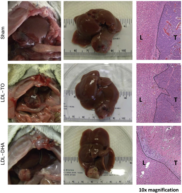
Foto: Therapeutic effect of hepatic artery injection (HAI) of LDL−DHA in HCC-bearing rats. In situ (left) and excised (middle) view of liver and H4IIE HCC tumor 3 days after sham or hepatic artery injection (HAI) with 2 mg/kg LDL nanoparticles. The right panel shows the corresponding histology at the liver tumor interface (10× magnification). Dotted line indicates liver–tumor boundary. L, liver; T, tumor.
Dit is heel verkort de aanpak die de onderzoekers toepasten:
- De onderzoekers gaven ratten met levertumoren of nanodeeltjes met LDL plus DHA of als controlegroep nanodeeltjes met LDL maar zonder DHA.
- Al na drie dagen waren de tumoren in de controlegroep gegroeid en toonden een goede bloedtoevoer (goed voor tumorgroei). Daarentegen toonden de ratten die LDL-DHA hadden gehad kleinere en bleke tumoren nagenoeg zonder bloedtoevoer (uiteindelijk kan de tumor niet overleven zonder bloedtoevoer).
- Meer dan 80% van de tumoren in de ratten die LDL plus DHA hadden gekregen was stervende.
De onderzoekers verklaren het uitstekende effect van LDL plus DHA als het effect van het Paard van Troje. Het is algemeen bekend, zo stellen de onderzoekers, dat tumorcellen gretig LDL opnemen om hun membraan (buitenkant van de cel) te verstevigen. Wanneer de nanodeeltjes dus bij de tumorcellen komen worden deze gretig opgegeten als het ware. Echter de tumorcellen weten niet dat er ook DHA bij zit en dat is volens de onderzoekers dodelijk voor tumorcellen. In feite wordt de tumorcel van binnenuit gedood door de DHA (omega-3).
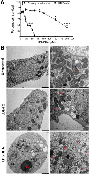
Foto:
Effects of LDL nanoparticles on primary hepatocytes and H4IIE cells. (A) MTS (3-(4,5-dimethylthiazol-2-yl)-5-(3-carboxymethoxyphenyl)-2-(4-sulfophenyl)-2H-tetrazolium) dose response assay of ACI rat primary hepatocytes and H4IIE cells to LDL−DHA (0−200 μM). Experiments were performed in triplicate wells with at least 3 independent runs. Results are expressed as mean ± SEM. ∗P ≤ .05; ∗∗∗∗P ≤ .0001 vs corresponding untreated group. All readings after 25 μM are significant at P ≤ .0001 for the H4IIE cells. (B) Transmission electron microscopy of H4IIE cells at baseline (untreated) and those treated with LDL−TO and LDL−DHA for 24 hours. Left panel: 2000 nm magnification bar, right panel: 1000 nm magnification bar. CC, nuclear chromatin condensation; L, lysosome; M, mitochondria. Arrowhead denotes swollen dysmorphic mitochondria; arrow condensed mitochondria; ×, autophagosome/autolysosome.
In het volledige studierapport dat gratis is in te zien: Hepatic Arterial Infusion of Low-Density Lipoprotein Docosahexaenoic Acid Nanoparticles Selectively Disrupts Redox Balance in Hepatoma Cells and Reduces Growth of Orthotopic Liver Tumors in Rats wordt uitvoerig deze studie beschreven en toegelicht. Misschien niet voor leken, maar zeker voor artsen en wetenschappers is dit studierapport uitstekend te begrijpen.
Hier het abstract van de studie:
An experimental nanoparticle therapy that combines low-density lipoproteins (LDL) and fish oil preferentially kills primary liver cancer cells without harming healthy cells
Hepatic Arterial Infusion of Low-Density Lipoprotein Docosahexaenoic Acid Nanoparticles Selectively Disrupts Redox Balance in Hepatoma Cells and Reduces Growth of Orthotopic Liver Tumors in Rats


 Article Info
Background & Aims
Dietary intake of the natural omega-3 fatty acid docosahexaenoic acid (DHA) has been implicated in protecting patients with viral hepatitis B or C from developing hepatocellular carcinoma (HCC). Little is known about the effects of DHA on established solid tumors. Here we describe a low-density lipoprotein−based nanoparticle that acts as a transporter for unesterified DHA (LDL−DHA) and demonstrates selective cytotoxicity toward HCC cells. We investigated the ability of LDL−DHA to reduce growth of orthotopic hepatomas in rats.
Methods
AxC-Irish (ACI) rats were given intrahepatic injections of rat hepatoma cells (H4IIE); 24 tumor-bearing rats (mean tumor diameter, ∼1 cm) were subject to a single hepatic artery injection of LDL nanoparticles (2 mg/kg) loaded with DHA (LDL−DHA), triolein (LDL−TO), or sham surgery controls. Tumor growth was measured by magnetic resonance imaging and other methods; tumor, liver, and serum samples were collected and assessed by histochemical, immunofluorescence, biochemical, and immunoblot analyses.
Results
Three days after administration of LDL−TO or sham surgery, the control rats had large, highly vascularized tumors that contained proliferating cells. However, rats given LDL−DHA had smaller, pale tumors that were devoid of vascular supply and >80% of the tumor tissue was necrotic. Four to 6 days after injection of LDL−DHA, the tumors were 3-fold smaller than those of control rats. The liver tissue that surrounded the tumors showed no histologic or biochemical evidence of injury. Injection of LDL−DHA into the hepatic artery of rats selectively deregulated redox reactions in tumor tissues by increasing levels of reactive oxygen species and lipid peroxidation, depleting and oxidizing glutathione and nicotinamide adenine dinucleotide phosphate, and significantly down-regulating the antioxidant enzyme glutathione peroxidase-4. Remarkably, the redox balance in the surrounding liver was not disrupted.
Conclusion
LDL−DHA nanoparticle selectively kills hepatoma cells and reduces growth of orthotopic liver tumors in rats. It induces tumor-specific necrosis by selectively disrupting redox balance within the cancer cell.
Gerelateerde artikelen
- RadboudUMC begint met long read whole genome sequencing testen voor opsporen van genetische afwijkingen bij zeldzame aandoeningen
- Grote verschillen in kwaliteit van zorg voor kankerpatienten tussen Nederlandse ziekenhuizen blijkt uit recent rapport van het NFK - Nederlandse Federatie van Kankerpatiëntenorganisaties
- CIPD = chronische inflammatoire demyeliniserende polyradiculoneuropathie geeft het beste resultaat met maximaal drie lage doses IVIg = Intraveneus immunoglobuline
- Lipidenverlagende medicijnen (LLM) (statines) hebben gunstige invloed op overall overleving van kankerpatienten met borstkanker, darmkanker en melanomen.
- Olanzapine, een atypisch antipsychoticum, blijkt door onbekende oorzaak veroorzaakte misselijkheid en braken bijna volledig te voorkomen en weg te nemen bij kankerpatienten met gevorderde kanker
- Oudere mensen met kanker lopen groter risico op de bacterie Clostridium difficile (veroorzaakt diarree) en overlijden daaraan ook vaker dan mensen zonder kanker.
- Opereren zonder snijden met bv. TACE, RFA, Nanoknife, yttrium-90, cryoablatie enz.: doe een consult bij specialistisch team in Nederland voordat u naar het buitenland op zoek gaat.
- Alle abstracten van SGO 2019 (50th Annual Meeting of the Society of Gynecologic Oncology)
- Slechts 5 van de 16 medicijnen, waaronder anti-PD medicijnen, goedgekeurd door FDA voor gebruik bij vormen van spijsverteringskanker blijken daadwerkelijk effectief in overall overleving, kwaliteit van leven en kosteneffectief.
- Vermagering door kanker en behandelingen van kanker is steeds beter tegen te gaan en verbetert vaker kwaliteit van leven en overall overleving. Een overzicht van effectieve behandelingen
- FDG PET/MRI geeft vergelijkbare of zelfs betere resultaten vergeleken met een FDG PET/CT bij verschillende vormen van kanker aldus een meta-analyse van 29 studies
- Kanker bij kinderen heeft lang na genezing nog gevolgen. 40 procent krijgt een levensbedreigende ziekte in haar/zijn verdere leven.
- LDL + DHA (visolie) verpakt en ingebracht met nanodeeltjes doodt razendsnel 80 procent van levertumoren en spaart gewone cellen blijkt uit dierstudie
- Radioloog Dr. Jelle Barentsz geeft uitleg via videofilmpjes over nieuwste niet-invasieve behandelingen en de rol van de beeldvorming via mp-MRI bij o.a. prostaatkanker.
- Voorlichtingsfolder van het KWF over hoe uitzaaiingen ontstaan
- Overlevenden van kinderkanker lopen 5 keer zo groot risico op ernstige endocriene - hormonale - aandoeningen op latere leeftijd
- Remming van glucose opname in senescente cellen - verouderde slapende stamcellen - doet tumorcellen afsterven en kan kankerpatienten behoeden voor een recidief
- Gerichte behandelingen op basis van moleculaire tumor profielen van de individuele kankerpatient lijken de toekomst te zijn. Aldus eerste gerandomiseerde SHIVA studie
- Algemeen: Studies, ook fase III studies, met een negatief of geen resultaat worden zelden of nooit gepubliceerd.
- Algemeen: Nieuw ontdekte genen - Fanconi genen - zou ontstaan van erfelijke kanker verklaren, aldus Nederlandse onderzoekers.
- Door bedrijven gesponsorde studies geven significant vaker een rooskleuriger beeld van de resultaten dan onafhankelijk uitgevoerde studies
- Algemene artikelen die met kanker te maken hebben maar niet specifiek bij een vorm van kanker
Plaats een reactie ...
1 Reactie op "LDL + DHA (visolie) verpakt en ingebracht met nanodeeltjes doodt razendsnel 80 procent van levertumoren en spaart gewone cellen blijkt uit dierstudie"